Patients Education
Your Trusted Partner in Cardiac Care
Understanding A Healthy Heart

The Heart’s Structure
Think of your heart as a powerful pump, divided into four rooms called chambers. The top two chambers are called atria, and the bottom two are called ventricles. The chambers work together, contracting and relaxing, to push blood through your body, like a well-coordinated team in a relay race.
Blood Flow
Blood from the body, filled with carbon dioxide and waste, enters the right atrium. This “used” blood is then sent to the right ventricle, which pumps it to the lungs. In the lungs, the blood picks up oxygen and releases carbon dioxide. The refreshed, oxygen-rich blood returns to the left atrium and is sent to the left ventricle. Finally, the left ventricle pumps the oxygen-rich blood out to the entire body, providing the fuel needed for every cell in the human body to function.


The Heart’s Electrical System
Your heart has its own electrical system that controls its rhythm. Picture it like a tiny conductor, orchestrating the beating of the heart. This electrical system sends signals through the heart muscle, telling it when to contract and relax. In a healthy heart, these signals create a steady, coordinated heartbeat, keeping everything in harmony. On the other hand, “arrhythmia” is when there is a disruption in the normal rhythmic cadence of the heart beat because of a break-down in the electrical system. “Electrophysiology” is the study of arrhythmia, and an electrophysiologist is a cardiologist with additional specialized training in treating arrhythmia.
The Heartbeat
Each heartbeat has two main parts: contraction and relaxation. During the contraction phase, called systole, the heart squeezes to send blood out to your body through the aorta–the body’s largest artery, and the source of all blood flow to the body. During the relaxation phase, called diastole, the heart fills with blood, like a sponge soaking up water. The rhythmic alternation of contraction and relaxation is what causes blood to circulate throughout the body. Damage to either the electrical system or the pump will cause the normal flow of blood to be in jeopardy.

Heart Health 101: The Basics of Maintaining a Healthy Heart
Eat a balanced diet
A heart-healthy diet can significantly reduce the risk of heart disease. Focus on including a variety of fruits, vegetables, whole grains, lean protein, and healthy fats in your daily meals. The American Heart Association recommends:
- Consuming at least five servings of fruits and vegetables daily.
- Choosing whole grains, which are higher in fiber and nutrients, over refined grains.
- Opting for lean protein sources like poultry, fish, beans, and low-fat dairy products.
- Incorporating healthy fats such as olive oil, avocados, and nuts, while minimizing saturated and trans fats.
Exercise regularly
Physical activity is crucial for a strong heart. The American Heart Association advises at least 150 minutes of moderate-intensity aerobic exercise or 75 minutes of vigorous aerobic exercise per week. This can include activities like brisk walking, swimming, cycling, or dancing.
Maintain a healthy weight
Carrying excess weight can strain your heart and increase the risk of heart disease. By combining a balanced diet with regular exercise, you can work towards achieving and maintaining a healthy weight. A body mass index (BMI) within the range of 18.5 to 24.9 is generally considered healthy.
Manage stress
Chronic stress can negatively impact heart health by raising blood pressure and contributing to unhealthy habits such as smoking, overeating, or excessive alcohol consumption. Develop stress management techniques like deep breathing exercises, yoga, meditation, or engaging in hobbies that help you relax.
Don’t smoke
Smoking is a major risk factor for heart disease. Quitting smoking significantly reduces the risk of heart attack and stroke. According to the American Heart Association, one year after quitting, the risk of coronary heart disease is cut in half, and within 15 years, it’s similar to that of a non-smoker.
Limit alcohol consumption
Excessive alcohol intake can lead to high blood pressure, heart failure, and increased calorie intake. The American Heart Association recommends limiting alcohol intake to one drink per day for women and up to two drinks per day for men.

Monitor Your Blood Pressure
Hypertension, commonly known as high blood pressure, exerts a significant impact on the development and progression of heart disease. The consistent elevation of blood pressure puts strain on the walls of arteries, leading to their thickening and narrowing. Over time, this condition, known as atherosclerosis, restricts blood flow to the heart muscle, depriving it of vital oxygen and nutrients. Additionally, the increased workload on the heart caused by hypertension can weaken the organ and lead to various cardiac complications such as heart failure, coronary artery disease, and arrhythmias. Furthermore, hypertension accelerates the formation of blood clots, increasing the risk of heart attacks and strokes. Given these detrimental effects, effective management of hypertension through lifestyle modifications and medication is crucial in reducing the incidence and severity of heart disease
Practical Tip: How to monitor Blood Pressure
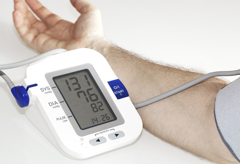
First, you will want to be sure that the blood pressure device you are using is accurate. A good way to do this is to bring it with you to your physician office visit and confirm that you get close (within 10mmHg) to the same result as your physician does. Second, you will want to make sure that the blood pressure result you get is the same (within 10mmHg) when measured between the right and the left arm. Some people will have a narrowing of blood flow to one or the other arm, resulting in a consistent blood pressure difference. If this is the case for you, the higher blood pressure is the correct number—you should always use the arm with the higher blood pressure for taking measurements.
Next, you will have to account for the fact that there may be natural variation in your blood pressure throughout the day. For some people the blood pressure is highest in the morning; for others it may be highest in the evening. The time that you take your blood pressure medication will affect this as well. So, if you really want to understand your blood pressure—for example, when you are adjusting blood pressure medication—you will need to check your blood pressure 3 or 4 times throughout the day. I often recommend taking the blood pressure in the morning when you wake up, at lunch, at dinner, and just before bed. Record these numbers in a spreadsheet for a week, and you will have a good sense for how your blood pressure fluctuates during the day. Of course, it is important to consistently take your regular blood pressure medication at the same times every day in order to get a true sense for your blood pressure, since changing timing of medication will affect your results.
A strategy of adjusting blood pressure medication on a daily basis according to measured blood pressure that day can be very tricky to navigate, since the medication takes an uncertain amount of time to be absorbed and take effect, and the blood levels of the medicine are affected by how regularly you take it. Therefore the goal should be to be taking the same medication each day, and not to vary it. If you track your blood pressure clearly in the manner described above, over the course of a few office visits you can generally arrive at a medication regimen that provides good blood pressure control throughout the day.
Finally, as adults get older, our kidneys become less effective at eliminating sodium (salt). This means that if you take a lot of salt in your diet, it can accumulate within the body, which in turn causes fluid to be retained, and raises blood pressure. So, if high blood pressure is an issue, you will want to restrict sodium in your diet. The American Heart Association website has extensive information for patients on diet and lifestyle, and is an excellent resource. It can be accessed by CLICKING HERE.
Blood Pressure Tracking
Managing blood pressure is key to maintaining cardiovascular health. High blood pressure puts hemodynamic stress on your arteries—including the arteries in your heart and brain—making them more prone to rupture or plaque build-up, resulting in heart attack, aneurysm, and stroke. The American Heart Association has extensive patient information about hypertension, which we encourage you to read.

Things get even more complicated if you are taking BP medication, because the effect of taking that medication takes some time to kick in, and then gradually phases in, and then gradually phases out, according to the speed of absorption from the GI tract and the drug half-life. For this reason, it is important to be “scientific” in your approach to blood pressure tracking, so that the information you collect can be properly understood by your physician, and correct decisions made about medication. We encourage you to take your blood pressure at specific times spread out across the day—for example:
- When you first wake up
- At noon or lunch time
- At dinner time
- At bed-time
Because blood pressure medication (if you take it) will impact your readings, we encourage you to take your medication at exactly the same time every day while you are tracking blood pressure. That will allow the doctor to understand the temporal relationship between medication and blood pressure. A sample work-sheet for tracking your blood pressure can be downloaded here.
You will want to maintain a reasonably stable diet in terms of salt-intake while doing this, since extra salt intake can increase your blood pressure through retaining fluid in the body. Another thing to consider is that people can develop narrowing of the arteries that supply the arms, which could lead to a falsely low BP reading. Therefore, at least once you should check the blood pressure in both arms to see if you have a significant BP difference (greater than 10mmHg) between arms. If you do, the higher BP is the correct one, and the one you should be tracking.

Blood Pressure Monitoring at Home
When it comes to tracking your blood pressure at home, it is essential to adopt a methodical approach that accounts for fluctuations in your blood pressure throughout the day and various factors influencing blood pressure—including when you take your blood pressure medication. The effectiveness of blood pressure medication is not always easy to determine because it may take hours for the drug to be absorbed and reach peak efficacy….and by that time your blood pressure may be coming down on it’s own due to normal fluctuation.
Below is an example of a well kept blood pressure table and medication record to use as a guide.
| Time | 08:00 AM | 12:00 PM | 05:00 PM | 10:00 PM |
| 01/05/23 | 120/80 | 128/82 | 115/78 | 142/88 |
| 01/06/23 | 118/75 | 130/88 | 120/80 | 160/93 |
| 01/06/23 | 125/85 | 128/85 | 130/86 | 165/95 |
Blood Pressure Medication:
8am: HCTZ 25mg; Metoprolol 50mg
2pm: Metoprolol 50mg
10pm: Lisinopril 5mg
Know Your Numbers
We encourage you to be an active participant in your health care. Regular check-ups with your healthcare provider can help you stay informed about your cholesterol, blood pressure, and blood sugar levels. Cardiology in particular is a field heavily influenced by data acquired through various testing means, and it is good to be familiar with these. One of the most important numbers to know is your ejection fraction (EF) which is an index of how strong your heart muscle is pumping. A normal EF is over 60%.
Our goal is to educate and guide patients through the process of understanding and managing their heart condition.


What’s The Difference Between Electrophysiology (EP) and Cardiology?
There are two sides to the heart—and also to heart disease. The side everyone sees first is the heart as a muscle, or the engine that pumps blood through the body. Damage to the engine will obviously impair its ability to perform its normal task. However, just like any engine, there is internal electronic circuitry that tells it when and how to work. Without that internal electronics, any motor would be useless. The heart is the same way—the heart muscle is the pump, but the internal wiring of that muscle, and the flow of electrical current through it, regulates the heart rhythms and rate. Because the electrical wiring is part of the heart muscle, damage to one is often associated with damage to the other.
SVT: Supraventricular Tachycardia

What is SVT?
Supraventricular tachycardia (SVT) is a heart rhythm disorder characterized by rapid heartbeats. Imagine your heart as a house with an electrical system. Normally, the electricity flows in an organized manner, keeping the lights on and appliances running smoothly. In SVT, a glitch occurs in the heart’s electrical system, causing it to go very fast at times when it shouldn’t. SVT starts abruptly, out of the blue, and seemingly for no reason.
Causes of SVT
There are several major types of SVT. In some cases SVT is caused by a collection of cells that become hyper-excitable, and fire very fast, causing the rest of the heart to follow along with it. These are called “automatic tachycardias”. If you drink too much coffee and your heart races, that is a form of automatic tachycardia. Not all automatic tachycardias are as benign as drinking too much coffee, however.
Another very common cause of SVT is caused by a “short-circuit” in the heart rhythm. Many people are born with an extra electrical fiber that connects the upper and lower chambers of the heart. While it may seem good to have a “spare” fiber just in case, it turns out to be a bad thing when the extra-fiber short-circuits the normal flow of electricity, causing very fast currents in the heart that generate the fast heart rate.

Symptoms of SVT
Patients are all different in their perception of SVT. Some may experience mild symptoms, while others may be extremely symptomatic, or even pass out. However, generally speaking, the following symptoms are often associated with SVT:
- Rapid, pounding heart beat (palpitation)
- Very strong heart beats
- Irregular heart beats, or a disruption of normal rhythm
- Dizziness, lightheadedness
- Fainting
- Shortness of breath
- Chest pain
- Feeling of anxiety or restlessness
- Throbbing sensation in neck or jaw
- Poking sensation in the left chest
These symptoms often start abruptly, out of the blue, and may last for minutes to hours.
Treating SVT
Prevention / Lifestyle changes: Avoiding triggers like stress, caffeine, and alcohol may reduce the likelihood of SVT for some people. In certain cases the SVT may be terminated by maneuvers that patients can be taught to perform, such as bearing down or exhaling against a closed mouth.
Medications: Some drugs can help regulate the heart’s electrical system. While these medications can reduce the tendency to go into SVT, there is a small risk of side effects, and it also does not fix the underlying problem that is causing the SVT. Young patients may not be receptive to taking medication the rest of their lives, while older patients who are already taking multiple medications may not mind as much.
Catheter ablation: In this procedure, an electrophysiologist inserts a small tube into a vein and threads it to the heart in order to locate the exact location of the electrical problem or short circuit. Once the problem is found, the abnormal tissue or circuit can be eliminated by applying heat to the tip of the catheter (called “ablation”) in order to cauterize the problem area. The most common causes for SVT have success rates for ablation in excess of 90-95% with a low risk of complication (1-5%). For many patients this risk compares favorably to the alternative of long term medication use.
AFib: Atrial Fibrillation
What is AFib?
Atrial fibrillation, or AFib, is a common heart rhythm disorder where the heart’s upper chambers, called atria, beat irregularly and rapidly, leading to poor circulation of blood within the heart itself, and impaired output of blood to the rest of the body. Imagine your heart as a symphony of many individual musicians coordinated by a conductor. In AFib, the musicians are no longer looking at the conductor, and each playing to their own wishes and completely out of sync with the rest of the musicians. This is why the most characteristic feature of AFib is an irregular pulse.

AFib is classified according to how long a patient has had it, and whether or not they are always out of rhythm. “Paroxysmal” AFib refers to patients who go in and out of AFib on their own. These patients generally have AFib episodes under 24-48 hours long, and then spontaneously return to normal rhythm. “Persistent” AFib refers to patients who remain in AFib once they go out of rhythm. These patients generally need a medical procedure (such as an external shock, or cardioversion) to reset the heart rhythm. “Permanent” AFib refers to patients who have been in AFib for so long (often years) that enough damage has been done to the upper chambers of the heart such that normal heart rhythm can no longer be restored. Patients generally progress, over a period of years, from paroxysmal to persistent, and finally to permanent AFib. During this time, progressively more injury is occurring to the atria as a result of the AFib, until a threshold is reached where normal rhythm is no longer possible. For this reason it is advantageous to intervene as early as possible when AFib is discovered.
The symptoms of AFib overlap with SVT: palpitations, heart pounding, irregular heart rhythm, dizziness, shortness of breath, chest pain. However, compared with SVT, very often the symptoms of AFib are more subtle, or patients may have no symptoms at all, particularly after starting heart medication. This is because symptoms are more associated with heart rate (too fast or too slow) and not so much with how regular (or irregular) the heart rhythm is. Patients with AFib on heart medication can have heart rates that are relatively normal, but still be out of rhythm. Importantly, though, the absence of symptoms does not mean the absence of complications (like stroke) or damage to the heart. Because symptoms may or may not be present, usually some form of cardiac monitoring will need to be done to know whether a patient is still having AFib.

Risks and Complications from AFib
The most important complication from AFib is stroke. Among all patients with stroke, AFib is among the most frequent causes. Unfortunately, too often the diagnosis of AFib is not known until the patient has already suffered from stroke. This has to do with the non-specific and often subtle symptoms from AFib. Certain individuals with AFib are at particularly high risk of stroke—these include those with existing heart disease of heart failure, hypertension, diabetes, vascular disease or prior stroke (or mini-strokes, also called TIA or Transient Ischemic Attacks). Older patients are also at increased risk. The risk of stroke can be reduced (but not eliminated) by taking powerful blood thinners, called “anticoagulants”. It is important to note that aspirin is not considered as an anticoagulant—this is a common misunderstanding.
In addition to stroke, AFib can cause symptoms of heart failure (shortness of breath, fatigue, poor exercise tolerance, swelling) as a result of impaired cardiac output. Patients with co-existing coronary artery disease may experience angina—heart pain from insufficient blood flow to the heart. Often quality of life is significantly impaired in patients with AFib, especially early on before patients learn to adapt to their condition.
Treating AFib
Treatment of Afib can be classified into a few major categories.
Anticoagulants: Blood thinners are the key first step in patients with AFib who have risk factors for stroke. This is because stroke is the most devastating complication from AFib. While blood thinners carry their own risks (of bleeding) for many patients this risk is less than the risk of stroke. An analysis and balancing of the risk of stroke versus risk of bleeding is an important part of the clinical evaluation of patients with AFib.
Medications: Prescription drugs can help control your heart rate and rhythm. Rhythm and rate are related but different features of normal heartbeat, and some medications address heart rhythm while others may address heart rate. While good heart rate control can usually be achieved with medication, keeping the heart rhythm in check is a much more difficult task. For patients with AFib, heart rhythm medication (called antiarrhythmic drugs) are only 40-60% effective. In addition, all antiarrhythmic drugs have potential for potentially serious complications, which mandates careful follow-up for anyone on long term antiarrhythmic drug therapy. However, for the right patient, antiarrhythmic drug treatment is both effective and well tolerated.
Catheter Ablation: Catheter ablation is a procedure performed by an electrophysiologist in which tubes are threaded through veins to the heart guided by X-ray. Using these catheters, carefully placed burns (“ablation”) are made at targeted regions in the heart around structured called the ”pulmonary veins”. In many patients the source of Afib resides within the pulmonary veins, and cauterizing around the pulmonary veins will block these abnormal impulses so they cannot escape into the heart to trigger AFib. Success rates for AFib ablation are about 85% if done early, before permanent damage has been done to the heart cells from long term exposure to AFib. If patients have been in constant AFib for years, the success rates may drop to 50-60%, or less. Clinical studies show that ablation is significantly more effective than antiarrhythmic medication at maintaining normal rhythm, and this often leads to better clinical outcomes. However, this benefit should be weighed against the small but finite risk of significant procedural complication, which is in the range of 1-5%. In addition, some patients will require a second procedure in order to achieve a lasting cure.
Surgical MAZE operation: In this procedure, a cardiac surgeon accesses the heart through the chest wall and makes cuts in specific regions of the atria in order to “channel” electrical currents in a normal manner. These procedures are the most effective at restoring normal rhythm, but are seldom the first choice of treatment because of the invasiveness of the procedure. However, more limited and less invasive MAZE operations have been developed that are often done in combination with catheter ablation. These so-called “hybrid” approaches are appealing options for patients with more advanced forms of AFib.
Device Implantation
In addition to catheter ablation, Dr Chow has exceptional expertise in cardiac device implantation. These devices include pacemakers, implantable cardioverter-defibrillators (ICDs), and cardiac resynchronization therapy (CRT) devices. The common purpose of all implantable cardiac devices is to restore and maintain a normal heart rate, and thereby improve symptoms, quality of life, and prevent complications like fainting, cardiac arrest or heart failure.

Pacemakers
A pacemaker is a small device implanted under the skin, usually in the region of the left upper chest near the collarbone. The device sits under the skin but above the muscle, and has wires (leads) that are threaded through veins down to the heart. When the heart beats too slowly, the pacemaker sends electrical signals to stimulate the heart to beat faster, at a normal rate. Pacemakers can sense patient movement, and during periods of high physical activity the pacemaker will know to pace the heart slightly faster to account for increased patient need for blood flow. In this way modern pacemakers are very “smart”. They pace when they are needed, and stop pacing when they are not needed. Pacemaker implantation is a relatively minor procedure that in the hands of an experienced operator will take under 30 minutes, and does not require an overnight hospital stay. Battery longevity is about 10 years.
Implantable Cardioverter-Defibrillators (ICDs)
An ICD looks very similar to a pacemaker, and is implanted using a similar technique. In fact, every ICD is also a pacemaker—but with a very important added feature. ICDs are able to treat cardiac arrest by either low voltage (rapid cardiac pacing) or high voltage (shock) methods. While no-one looks forward to an ICD shock, the key point to remember is that shocks are only intended to be delivered in the event of a life-threatening heart arrhythmia (cardiac arrest). Clinical studies show that for high risk patients (especially with an EF < 35%) patient survival is significantly superior with ICD treatment as compared with medical therapy alon. ICD implantation is a relatively minor and safe surgical procedure that can be done in about 30 minutes, and does not require an overnight hospital stay. Both pacemakers and ICDs are often implanted in outpatient surgery centers.
Cardiac Resynchronization Therapy (CRT) Devices
CRT devices are also called “bi-ventricular pacemakers” and “bi-ventricular ICDs”. These devices can be considered as “regular” pacemakers and ICDs with one important additional component—a left ventricular lead. During the implant process an additional pacing wire is threaded through a vein in the heart that leads to the surface of the left heart chamber (left ventricle). By pacing the left ventricle simultaneously with the right heart chambers, these sophisticated devices improve the strength and efficiency of the heart beat. Over time, these devices can result in strengthening and healing of damaged heart muscle. Bi-ventricular devices are commonly used in patients with symptoms of congestive heart failure (shortness of breath, swelling, fatigue) due to a weak heart muscle. These devices can significantly improve symptoms and daily function in patients limited by heart failure. A bi-ventricular device implant is an outpatient hospital procedure that takes about 2 hours to implant, has relatively low risk, and does not require an overnight hospital stay.
General Consultative Cardiology & Diagnostic Testing
Our practice offers comprehensive consultative services in all aspects of general cardiology and electrophysiology. We cover the broad spectrum of heart diseases, from preventative cardiology to chest pain and coronary artery disease, to palpitation and heart arrhythmia. Cardiology is a field in which modern technology has revolutionized our ability to understand and diagnose heart problems. Diagnostic studies and tests are commonly used together with the physician’s clinical evaluation to identify the specific problems.
Our practice offers diagnostic cardiac testing on site within our office. This makes scheduling easy, faster, and more convenient for patients. More importantly, it centralizes cardiac care and helps get to the cause of the patient’s heart problem quickly and with quality.

Common Diagnostic Tests

Electrocardiogram (ECG)
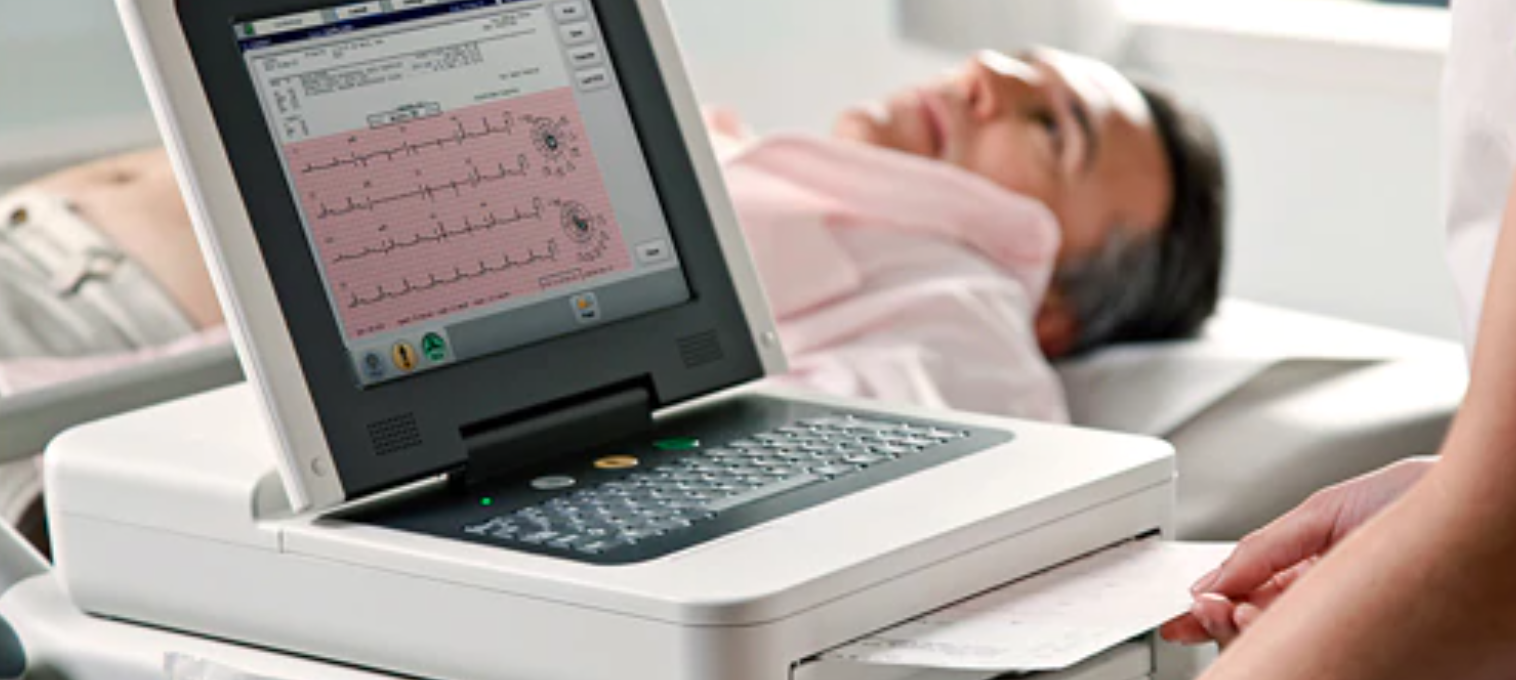
An ECG records your heart’s electrical activity. During this test, small sensors called electrodes are placed on your chest, arms, and legs. The ECG machine then measures and records the electrical signals generated by your heart as it beats, helping doctors identify any irregularities in your heart’s rhythm, as well as providing clues about other structural heart problems or damage that may have occurred in the past. However, the ECG in many cases is used as a screening tool, and more accurate and definitive cardiac tests exist and are discussed below.
Holter / MCOT Monitor
Holter monitoring and MCOT (Mobile Cardiac Outpatient Telemetry) monitoring are both diagnostic tests used to monitor and record the electrical activity of the heart over an extended period. These tests are particularly helpful in detecting and diagnosing abnormal heart rhythms or arrhythmias that may not be captured during a short-duration ECG.

Holter monitoring involves wearing a portable device called a Holter monitor, which records the heart’s electrical signals continuously for 24 to 48 hours or even longer. Modern devices are very small and non-intrusive. The patient is instructed to continue with their normal activities while wearing the device, and they may be asked to keep a diary to document any symptoms or activities that occur during the monitoring period. On the other hand, MCOT monitoring is a similar test that provides longer-term monitoring, for a week or longer.
By capturing and analyzing the heart’s electrical activity over an extended period, Holter monitoring and MCOT monitoring allow physicians to correlate symptoms with recorded events. This can help determine the underlying cause of symptoms such as palpitations, dizziness, or fainting spells. Based on the results, physicians can develop an appropriate treatment plan, which may include lifestyle modifications, medications, or further diagnostic tests.

Echocardiogram (ECHO)
Cardiac echocardiography, commonly referred to as a cardiac echo or echocardiogram, is a non-invasive imaging test that utilizes ultrasound waves to create real-time images of the heart. It provides valuable information about the structure and function of the heart, helping healthcare professionals evaluate a patient’s heart health and diagnose various cardiac conditions.
The results of a cardiac echo can help healthcare professionals diagnose and manage various cardiac conditions. It can assist in the diagnosis of conditions such as heart failure, coronary artery disease, cardiomyopathies, and heart valve abnormalities. The information obtained from the echo can guide treatment decisions, including the use of medications, lifestyle modifications, or the need for further diagnostic tests or interventions.
Cardiac echo is a safe and painless procedure that does not involve radiation exposure. It is widely used due to its ability to provide detailed and real-time images of the heart, aiding in the assessment of heart function and the diagnosis of cardiac abnormalities.
Carotid Ultrasound
Carotid ultrasound is a non-invasive imaging test that uses high-frequency sound waves to produce images of the carotid arteries, the blood vessels in the neck that supply the brain with oxygen-rich blood. During the procedure, the patient lies on their back, and a small handheld device called a transducer is placed on the neck. The transducer emits sound waves that bounce off the blood vessels and create images of the carotid arteries on a monitor.


Stress Test
A cardiac stress test, also known as an exercise stress test, is a diagnostic test that assesses the heart’s ability to function under physical stress. There are two main types of stress tests: treadmill stress testing and pharmacological stress testing (i.e. non-exercise stress testing). Treadmill stress testing involves walking on a treadmill while being monitored by a healthcare professional, whereas pharmacological stress testing is done with the use of medications that simulate the effects of exercise on the heart.
The results of a cardiac stress test can help diagnose significant coronary artery disease and assess the risk of heart attack. If the test shows normal results, it suggests that the patient’s heart is functioning properly and that there is no significant blockage in the coronary arteries. However, abnormal results indicate that there may be a serious blockage or narrowing of the coronary arteries, which reduces blood flow to the heart muscle and can lead to chest pain or a heart attack.
It is important to note that not all blockages affect blood flow in your coronary arteries. Blockages less than 70% are generally not flow-limiting, and can produce normal findings on stress test. However, because blood flow is normal in those cases, blockages under 70% are generally not treated invasively. Therefore stress testing is very useful for determining patients with severe blockages that require further testing or aggressive treatment, such as coronary angiography, coronary stenting or bypass surgery.
It is also important to note that stress testing provides a snapshot of your heart’s circulation at one point in time. That result is not static, and over time things may change. Results of stress testing are most reliable if done within the past year, and begin to gradually fade in reliability after that.
Advanced Cardiac Imaging Studies
a. CT scan (Computed Tomography): Coronary CT angiography (CTA) is a non-invasive imaging technique that provides detailed images of the coronary arteries. It involves the use of a computed tomography (CT) scanner and a contrast dye injected into the bloodstream to visualize the coronary arteries. The CT scanner rotates around the patient, capturing multiple X-ray images that are reconstructed into 3D images of the heart and its blood vessels.
The results of a coronary CTA can help determine the presence and severity of coronary artery disease (CAD). If the test shows normal or minimal plaque buildup in the coronary arteries, it suggests that the patient has a low risk of significant blockages and a lower risk of heart attack. On the other hand, if the test reveals significant plaque buildup, narrowing of the arteries, or blockages, it indicates a higher risk of heart attack and the need for further evaluation or treatment.
b. MRI (Magnetic Resonance Imaging): Cardiac magnetic resonance imaging (MRI) is a non-invasive imaging technique that provides detailed images of the heart’s structure and function. It utilizes a powerful magnetic field and radio waves to generate highly detailed images of the heart and its surrounding structures.
During a cardiac MRI, the patient lies on a table that slides into the MRI machine. The machine creates a strong magnetic field around the patient, and radio waves are directed towards the body. The interaction between the magnetic field and the radio waves generates signals that are detected by the MRI machine. These signals are processed by a computer to create high-resolution images of the heart.
Cardiac MRI can provide information about the heart’s size, shape, and function, as well as detect abnormalities in the heart muscle, valves, and blood vessels. It can assess cardiac function, including measurements of ejection fraction (the percentage of blood pumped out with each heartbeat), cardiac volumes, and blood flow patterns. It can also detect and characterize tissue damage, such as scar tissue resulting from a heart attack.
The detailed images obtained from a cardiac MRI can help diagnose various cardiac conditions, such as heart muscle diseases (cardiomyopathies), congenital heart defects, heart valve abnormalities, and tumors. It can also provide valuable information for treatment planning, monitoring disease progression, and evaluating the effectiveness of interventions.An MRI uses powerful magnets and radio waves to create detailed images of your heart’s structures and blood vessels.
Telehealth and Remote Monitoring of Cardiac Data
Telehealth refers to the use of digital communication technologies to provide healthcare services remotely. It allows patients to access medical care and consultation without the need for in-person visits to a healthcare facility. Through telehealth, patients can interact with healthcare providers using video calls, phone calls, or secure messaging platforms. This method of healthcare delivery offers numerous benefits, including improved access and convenience for patients.

Telehealth also potentially offers enhanced convenience for all patients. Instead of spending time commuting to and waiting at the doctor’s office, patients can schedule virtual appointments and wait in the comfort of their own home.
As part of our commitment to providing exceptional service, our cardiology practice offers telehealth services to patients. By utilizing telehealth, we aim to improve accessibility and convenience while maintaining the same quality of care. Through secure and confidential video consultations, we can discuss your cardiac health, review test results, answer your questions, and develop personalized treatment plans—all without you having to leave home.
Anyone who may have been exposed to COVID recently is asked to conduct their visit over telehealth.
Please reach out to our practice to learn more about our telehealth services and how we can assist you in managing your cardiac health effectively.
